MR measurement
We provide standard MR imaging (MRI) and MR spectroscopy (MRS) methods, as well as MR methods based on our methodological research for
- MR measurements of nonliving samples, e.g. for characterization of diagnostic and theranostic nanoparticles, geological and other materials,
- MR measurements of ex vivo tissue samples,
- Longitudinal in vivo MR measurements of mice and rats.
List of the currently provided methods:
Anatomy
MRI offers excellent soft-tissue contrast. We provide high-resolution cross-sectional images acquired in a variety of orientations and anatomical locations. We typically achieve in-plane resolutions of 50–100 µm for mouse brain imaging, 100–200 µm for whole-body or organ imaging, and down to 25–50 µm isotropic for ex vivo studies. Tissue contrast can be tailored by employing sequences weighted by T1, T2, T2*, or susceptibility (SWI). In addition, specialized techniques are available for the selective suppression of fat, water, or cerebrospinal fluid (CSF) signals, enhancing visibility of specific structures. MRI acquisition can be synchronized with cardiac motion (e.g., for heart imaging) and/or respiration, enabling the capture of cine sequences that visualize organ motion. Following the acquisition, post-processing pipelines can provide calculation of tissue volumes, which support quantitative analysis and provide robust metrics for comparison across experimental groups or longitudinal studies.
Relaxometry
Magnetic resonance relaxometry provides quantitative maps of relaxation times (T₁, T₂, and T₂*), offering a direct way to measure tissue properties rather than just visual contrast. These maps reflect aspects of the tissue microstructure. Because relaxation times are sensitive to pathological changes, relaxometry can be used to study:
- fibrosis and scarring in organs such as liver, heart, or kidney,
- efficacy of contrast agents and their distribution in tissues, this is used in our biodistribution studies of experimental contrast agents, targeted agents, and theranostics,
- neurodegenerative processes, where T₁ or T₂* changes may indicate iron accumulation or demyelination,
- tumor characterization and therapy monitoring, where altered tissue composition is reflected in relaxation properties,
The advantage of relaxometry is that it yields objective, quantitative values that are comparable across animals, experiments, and time points. This makes it particularly useful for longitudinal studies, group comparisons, and therapy evaluation.
 T1 map of Myocardium (ms)
Non-Contrast Angiography Using Time-of-Flight MRI
Our Magnetic Resonance Department offers non-invasive, non-contrast angiographic imaging using the Time-of-Flight (TOF) MRI technique.
This method enables detailed visualization of blood vessels without the need for contrast agents, making it ideal for longitudinal studies or situations where contrast administration is not suitable.
We specialize in small animal imaging, with optimized protocols for:
- Mouse and rat brain vasculature
- Other anatomical regions of interest (upon request)
Key features:
- High-resolution vascular imaging
- No contrast agents required
- Suitable for repeated measurements in longitudinal studies
- Tailored imaging protocols available on demand
- Experienced technical support and consultation
Typical applications include research into stroke (ischemic models), cerebrovascular diseases, vascular development, and preclinical drug testing.
Our TOF MRI method is particularly valuable in neuroscience and cardiovascular research, where precise, non-invasive imaging is essential.

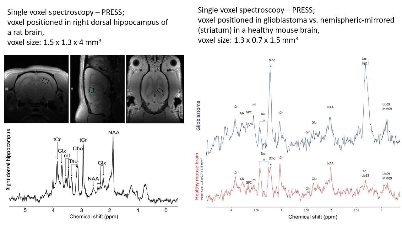
MR spectroscopy
We offer 1H MRS measurements on phantoms, a variety of samples (biological and non-biological), as well as in mouse and rat models in vivo. MRS allows non-invasive monitoring of metabolite levels, enabling researchers to observe metabolic changes, evaluate drug effects, and investigate biochemical alterations associated with disease. This versatile technique provides insights into energy metabolism, neurotransmitters, and other biochemical pathways, supporting preclinical research and drug development.
Using single voxel spectroscopy methods, we provide relative concentrations of the following metabolites:
total creatine (creatine and phosphocreatine)
total NAA (N-acetyl aspartate and N-acetyl aspartyl glutamate), known as a marker of functional neurons,
total choline (choline-containing compounds), higher levels suggest membrane turnover, while lower levels may indicate a lack of acetyl choline,
glutamate, an excitatory neurotransmitter,
glutamine, synthesized from glutamate in astroglia, a marker of astroglia,
Glx (sum of glutamate and glutamine) provides more precise results, as glutamate and glutamine have similar molecular structures
myo-inositol, proposed as a marker of glial cells, but detected in various cell types,
taurine, suggested as osmoregulator and modulator of neurotransmitter actions, neuroprotective potential,
lactate, the end-product of anaerobic glycolysis.
The brain structures of interest:
prefrontal cortex,
sensorimotor cortex (right and left),
striatum (right and left),
hippocampus (right and left),
thallamus (right and left),
cerebellum.
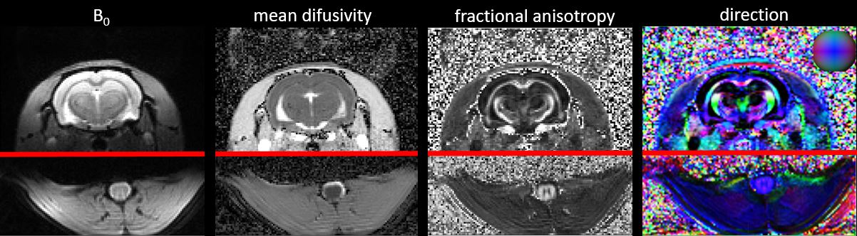
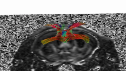
Diffusion-Weighted and Diffusion Tensor Imaging (DWI / DTI)
We offer advanced Diffusion-Weighted Imaging (DWI) and Diffusion Tensor Imaging (DTI) for a variety of small animal models, including rats, mice, and other species upon request.
These techniques allow us to investigate tissue microstructure properties related to water diffusion, providing valuable insights into both healthy and pathological states.
We routinely perform diffusion imaging of the:
- Brain
- Spinal cord
- Kidneys
From the acquired data, we generate a wide range of diffusion parameter maps, including:
- Mean Diffusivity (MD)
- Fractional Anisotropy (FA)
- Directionality (principal diffusion directions)
- Apparent Diffusion Coefficient (ADC)
- Trace
We also perform tracking of neuronal fibres (tractography) to reconstruct white matter pathways, particularly in the brain and spinal cord.
These methods are applicable to research in:
- Neurological diseases (e.g., stroke, neurodegeneration, trauma)
- Spinal cord injury and regeneration
- Renal disorders
- Preclinical testing and developmental studies
- Our team provides complete support, from protocol optimization to data acquisition and processing.
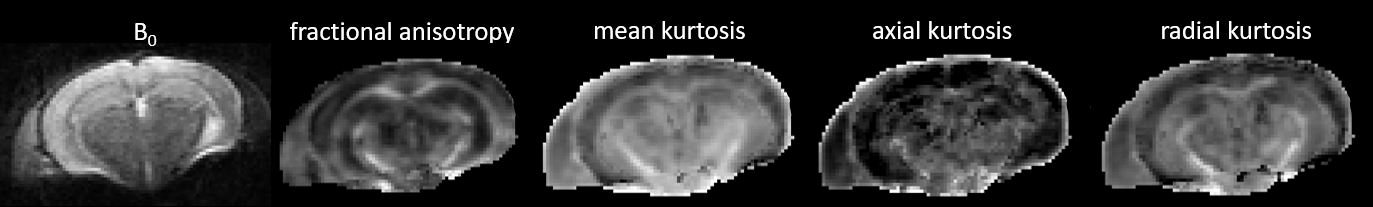
Diffusion Kurtosis Imaging (DKI)
We offer Diffusion Kurtosis Imaging (DKI) for rats, mice, and other small animal models, providing a deeper insight into tissue microarchitecture beyond what standard diffusion methods can reveal.
Unlike DTI, which assumes Gaussian water diffusion, DKI quantifies deviations from Gaussian diffusion, offering a more nuanced picture of tissue microstructure. This makes it a powerful tool for studying complex or heterogeneous biological tissues.
We routinely apply DKI to the:
- Brain
- Spinal cord
- Kidneys
DKI provides additional biomarkers of tissue integrity and pathology, useful in the detection and monitoring of various diseases, including:
- Neurodegenerative disorders (e.g., Alzheimer’s disease, Parkinson’s disease)
- Ischemic stroke and traumatic brain injury
- Spinal cord injury
- Renal dysfunction and fibrosis
By analyzing metrics such as mean kurtosis (MK), axial kurtosis (AK), and radial kurtosis (RK), DKI allows researchers to assess microstructural complexity, tissue heterogeneity, and cellularity more accurately than conventional DTI.
As with our other diffusion imaging services, we offer complete support from customized protocol design to data processing and interpretation.
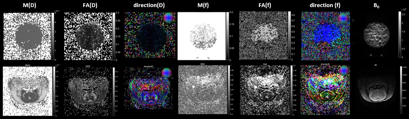
Intravoxel Incoherent Motion (IVIM) MRI
We offer Intravoxel Incoherent Motion (IVIM) MRI for small animal models, including mice, rats, and other species on request. This advanced technique represents an extension of DTI, adding valuable perfusion information to standard diffusion imaging.
Unlike conventional DTI, IVIM distinguishes between:
- True molecular diffusion (slow component)
- Pseudodiffusion caused by microvascular perfusion (fast component)
This dual insight allows researchers to assess both tissue microstructure and microvascular properties simultaneously.
We offer two types of IVIM imaging:
- Standard (single-direction) IVIM
- Multidirectional IVIM, enabling the estimation of:
- Perfusion and diffusion anisotropy
- Directionality of both diffusion and perfusion
This multidimensional approach offers a more comprehensive characterization of tissue and vascular microarchitecture, including:
- Orientation and structure of capillary networks
- Tissue integrity and heterogeneity
- Differentiation of pathological vs. healthy regions
IVIM MRI is particularly useful in studies of:
- Brain and spinal cord perfusion
- Renal microcirculation and pathology
- Tumor characterization and drug response
- Neurovascular disorders
As with our other MR techniques, we provide custom protocol development, data acquisition, and advanced post-processing tailored to your research needs.
Perfusion imaging - ASL
Arterial Spin Labeling (ASL) is an endogenous contrast method i.e. does not need contrast agent injection during MR measturement. Instead of intravenous application of contrast agent spins are labeled using RF pulses. Usually changes in signal intensity or T1 relaxation time in labeled and control images is observed. A quantitative tissue-specific map of perfusion can be determined. Primary applications are in brain, kidneys or heart.

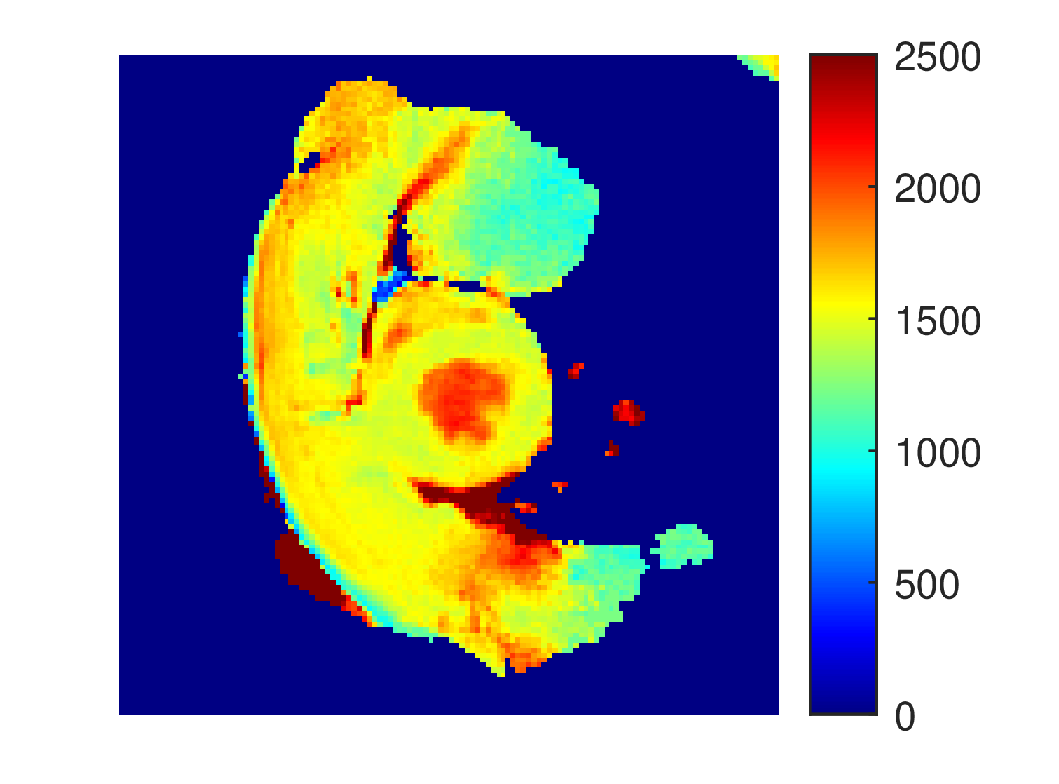
Kidney Flowmap
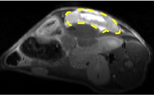
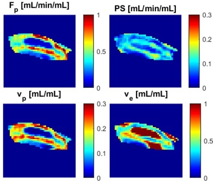
Perfusion imaging - DCE-MRI, DSC-MRI
Dynamic Contrast-Enhanced and Dynamic Susceptibility Contrast MRI (DCE-MRI and DSC-MRI) track how a contrast agent (commonly gadolinium-based) passes through tissue and blood vessels. These methods allow non-invasive evaluation of microvascular properties, which are crucial in studying tumor angiogenesis, therapy response, and vascular changes in organs such as brain, kidney, liver, and prostate. With these methods, maps of the following quantitative parameters can be obtained:
Blood flow – volume of blood delivered to tissue voxel per unit time (perfusion).
PS (permeability–surface area product) – direct measure of vascular-wall permeability.
Blood volume – fraction of tissue volume occupied by blood (vascular density).
Extracellular extravascular volume fraction – fraction of tissue volume outside vessels and cells.
Mean transit time – average time blood requires to traverse a tissue region (circulatory efficiency).
Ktrans – volume transfer constant; rate of contrast-agent transport into extravascular space.
Together, DCE-MRI and DSC-MRI provide a detailed picture of tissue vascularization and microenvironment. This makes them powerful tools in preclinical research on cancer, stroke, and other diseases where blood supply and vessel properties play a central role. It can also be used for assessing the permeability of the blood-brain barrier (BBB).